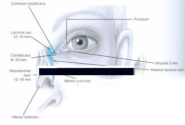
Tear duct surgery or Dacryocystorhinostomy (DCR surgery) involves the exploration and reconstruction of the tear ducts that normally drain tears from the ocular surface into the nose and mouth. Excessive tearing is a common complaint Dr. Zoumalan evaluates in his patients. Excessive tearing can result from a variety of causes and his role is to evaluate the tear duct system and to make sure your symptoms are not a direct result of a stenosis or obstruction within the tear duct system. The lacrimal system is also referred to as the nasolacrimal system, tear duct system, or lacrimal drainage apparatus. It is a drainage system that allows tears from the ocular surface to drain down into the nasopharynx. Patients can be present with problems within the lacrimal system which can result in a stenosis or a blockage anywhere throughout the passageway. Patients usually are present with tearing in that particular eye and may or may not have a history of recurrent infections. See below for an anatomic diagram of the nasolacrimal duct.
Diagram adapted from: Zoumalan, C.I, Olmos, A. Cockerham, K.P. Eyelids and Lacrimal System. In Agarwal A, ed.: Color Atlas of Ophthlamology, Second Edition: New York, NY: Thieme Medical Publishers; 2009.
The tears drain from two openings within the eyelids, each called a punctum, which are 1-2 millimeters in diameter. They each drain into a canaliculus, both of which meet to form a lacrimal sac. Often the puncta or canaliculi are stenotic or blocked and may require reconstruction procedure to open the lumen up if possible. The canaliculi then drain into the lacrimal sac, which then empties into the nasolacrimal duct within the nasal passageways.
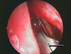
This image was taken intra-operatively during an endoscopic DCR surgery. It shows the new lacrimal system bypass that is created in order to allow the patient to appropriately drain their tears.
We often see obstruction around the lacrimal sac area as well; and a complete lacrimal system examination needs to be performed to identify the problematic area within the lacrimal system. Females and patients over the age of 50 are more at risk for developing a nasolacrimal duct obstruction. This is likely due to a variety of reasons including their thin nasolarimal ducts relative to males, and hormonal changes which may predispose females to increased bouts of inflammation or swelling of their nasolacrimal duct lining tissue. If there is an obstruction near the lacrimal sac and the patient has constant tearing and/or history of infections, a dacryocystorhinostomy (DCR) surgery may need to be performed. This is a surgery with very high success rates and allows for the creation of a bypass system for the tears to drain into the nasal passageways. Dr. Zoumalan specializes in performing DCRs and utilizes the latest innovations in DCR surgery often employing an endoscope for optimal visualization. In selected DCR cases, an endoscope can be used and patients may not require an external (or visible) incision.
Dacrycystorhinostomy: What is the difference between an endoscopic versus external DCR surgery?
Dacrycystorhinostomy: How long are the tubes left in place after tear duct surgery?
Dacrycystorhinostomy: What should you expect at your preoperative visit for your upcoming orbital surgery?
Dacrycystorhinostomy: What should you expect on the day of your orbital surgery?
Dacrycystorhinostomy: What should you expect after your orbital surgery in terms of recovery and pain control?
Dacrycystorhinostomy: What are the risks and complications to orbital surgery?
Dacrycystorhinostomy: Does insurance cover orbital surgery?
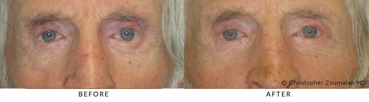
This patient had to undergo a tear duct reconstructive surgery to help his tears drain better in his left eye. He had a nasolacrimal duct obstruction. He underwent a dacryocystorhinostomy (DCR) surgery to create a new passageway to help his tears drain better. An external DCR was performed in the left eyelid and there was a small incision (6mm long) made along his lower eyelid skin. The post operative photograph was taken 2 months after surgery. There is no visible scar noticeable. Silicone tubes are left inside the tear ducts and they act as stents to keep the new tear duct patent during the healing process. They are removed usually three months after surgery. They are barely visible to observers since they are small and transparent.
Preoperative Diagnosis: Left lower lid entropion
Procedure performed: Left lower lid entropion repair
Disclaimer:
*Individual results may vary
All before and after pictures displayed are real patients who have consented to having their pictures published on our site. Individual results will vary with each patient and Dr. Christopher Zoumalan does not guarantee any outcomes of procedures shown. All pictures are meant for reference and illustrative purposes only.
Dr. Christopher Zoumalan is a board-certified oculoplastic surgeon in Beverly Hills, specializing in aesthetic and reconstructive oculoplastic surgery. Dr. Zoumalan is passionate about helping patients reach their ideal aesthetic and restore functionality with the leading cosmetic eyelid surgery, reconstructive eyelid surgery, and non-surgical cosmetic procedures. Dr. Zoumalan has been featured on several television shows and has traveled the world as a trusted oculoplastic specialist. Browse the video gallery below to learn more about oculoplastic procedures and treatments.